McGill
Mössbauer Group


Our 166Er Mössbauer system came on line in November 2001
This is our third cold-source Mössbauer isotope.
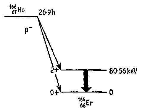
166Er Mössbauer uses the 80.56 keV level which is populated by the decay of 27 hour 166Ho. This isotope was prepared by neutron irradiation of 165Ho in the `SlowPoke' at Ecole Polytechnique, Montreal.


Some test data
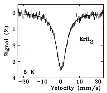
This was is our first spectrum.
The sample was ErH2
The source was 27 mCi of 166Ho in Ho0.4Y0.6H2.
The sample and source were both held at 5 K.
The velocity scale was determined using simultaneous calibration with a 57Co source and iron metal sample.

We obtained a linewidth (HWHM) of 3.30(1) mm/s.

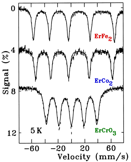
ErFe2 (top) and ErCo2 (centre), provide examples of magnetically split 166Er Mössbauer spectra. As both samples are fully ordered, relaxation effects are absent and sharp lines are observed (2.46(4) mm/s).

The asymmetry is due to a significant electric field gradient in addition to the 800 Tesla hyperfine field.

Both spectra were fitted with lineshapes calculated using the full Hamiltonian for this Mössbauer transition.

The bottom spectrum (ErCrO3) shows a smaller Er moment (the hyperfine field is only 500 Tesla), while the much broader lines (3.79(5) mm/s) combined with the form of the mis-fit, clearly indicate the presence of relaxation.


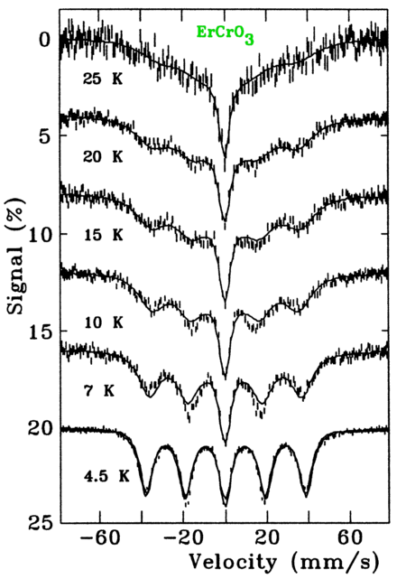
The temperature dependence of the 166Er Mössbauer spectra of ErCrO3 clearly show the influence of slow paramagnetic relaxation.

At 25 K the spectrum exhibits broad wings around the central line. As the sample is cooled, the fluctuations slow, and the wings resolve into distinct lines.

The spectra were fitted using a standard 2-state fluctuation model.

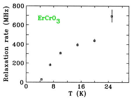
The result of fitting a relaxation model to the ErCrO3 spectra shows how the fluctuation rate grows rapidly with increasing temperature.


Example of application:
Er3Ge4
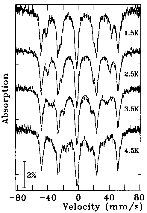
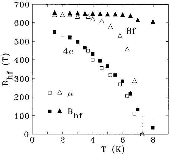
Erbium atoms occupy two crystallographically distinct sites in this material. Neutron diffraction studies (JMMM 169 (1997) 253) showed that the Er moments at these two sites were very different at 1.15(2):1 for the 8f and 4c sites respectively.

The 166Er Mössbauer spectra at the left clearly show the two pentets arising from the 8f and 4c Er sites (clearest at 2.5K), and give a hyperfine field ratio of 1.183(5), consistent with the earlier neutron diffraction work. The line broadening visible as the 4c pentet collapses on heating, is due to slow electronic relaxation.

The hyperfine field at the 4c site tracks the neutron diffraction derived moment extremely well, despite some complications due to slow electronic relaxation.

By contrast, relaxation at the8f site is so slow that we observe essentially no temperature dependence at all.

TN = 7.2K and is marked by the dotted vertical line.


Updated: 04/Aug/03
All images copyrighted by:
Dominic Ryan, ERP 425, (514) 398-6534